Mouse Bone Marrow Endothelial Cell Culture
| Mouse Endothelial Cell (mEC) Complete Media Recipe | |
| File Size: | 121 kb |
| File Type: | |
| Mouse Endothelial Cell (mEC) Culture Procedures | |
| File Size: | 73 kb |
| File Type: | |
| Mouse Endothelial Cell (mEC) Isolation Protocol | |
| File Size: | 155 kb |
| File Type: | |
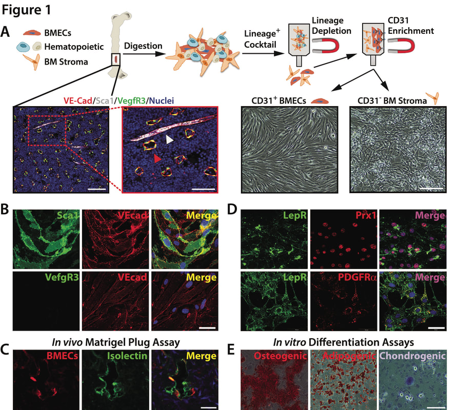
Generation of BM-Derived Endothelial and Stromal Cells (A) Schematic of BMEC and BMS cell isolations. VECAD+SCA1+VEGFR3− arteriole (white arrowhead) and VECAD+SCA1−VEGFR3+ sinusoidal (red arrowhead) BMECs in vivo (scale bar represents 100 and 50 μm). Phase-contrast images of isolated BMEC-Akt1 and BMS-Akt1 cultures (scale bar represents 200 μm). (B and C) BMEC-Akt1 cultures demonstrate VECAD+SCA1+VEGFR3−arteriole staining (B) (scale bar represents 50 μm) and produce vessels in vivo (C). BMEC-Akt1 (red), but not BMS-Akt1 (blue), co-localize with intravitally labeled vasculature (Isolectin-B4; green) in matrigel plugs (scale bar represents 25 μm). (D and E) BMS-Akt1 cultures display PDGFRA+PRX1+LEPR+ BMS staining (D) (scale bar represents 50 μm) and give rise to osteogenic (Alizarin Red S), adipogenic (Oil Red O), and chondrogenic (Toluidine Blue O) progeny in vitro (E) (scale bar represents 200 μm).
Poulos MG, Crowley MJP, Gutkin MC, Ramalingam P, Schachterle W, Thomas JL, Elemento O, Butler JM. Vascular Platform to Define Hematopoietic Stem Cell Factors and Enhance Regenerative Hematopoiesis. Stem Cell Reports. 2015 Nov 10;5(5):881-894. doi: 10.1016/j.stemcr.2015.08.018. Epub 2015 Oct 1. PMID: 26441307; PMCID: PMC4649106. https://pubmed.ncbi.nlm.nih.gov/26441307/


